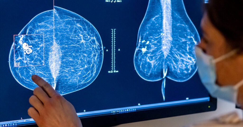
Clinics across the country are starting to offer patients a new service: having their mammograms read not only by a radiologist, but also by an artificial intelligence model. Hospitals and companies that provide these tools tout their ability to speed up the work of radiologists and detect cancer earlier than standard mammograms alone.
Currently, mammograms detect about 87 percent of breast cancers. Younger women and those with dense breasts are more likely to get cancer. They sometimes lead to false positives that require more tests to rule out cancer, and they can also show precancerous conditions that may never cause serious problems but still lead to treatment because the risk of not treating them can’t be predicted.
“It’s not a perfect science by any means,” said Dr. John Lewin, chief of breast imaging at Smilow Cancer Hospital and Yale Cancer Center.
Experts are excited about the prospect of improving the accuracy of screening for breast cancer, which diagnoses 300,000 women each year in the United States. But they also have concerns about whether these AI tools will work well in a wide range of patients and whether they can meaningfully improve breast cancer survival.
How does AI analysis work?
Mammograms contain a wealth of information about the breast tissues and ducts. Certain patterns, such as bright white spots with jagged edges, can be a sign of cancer. Thin white lines, by contrast, may indicate calcifications that may be benign or may need further testing. Other patterns may be difficult for people to differentiate from normal breast tissue.
AI models can, in some cases, “see what we can’t see,” said Dr. Katerina Dodelzon, a radiologist specializing in breast imaging at NewYork-Presbyterian/Weill Cornell Medical Center.
When an image is run through an AI program, the software highlights suspicious areas that require further attention from a radiologist. Some models can also score images to help busy radiologists prioritize which scans to review first.
“I easily read 100 screening mammograms in one day,” said Dr. Carolyn Malone, a breast radiologist at the John Theurer Cancer Center at Hackensack University Medical Center. “I can start reading what the AI says are more complex.”
In one of the largest AI mammography studies, a model used in Sweden improved breast cancer detection by 20%. In a trial involving 80,000 women, the software found six cases of cancer in every 1,000 women, while radiologists found five in every 1,000 women.
A 2022 Danish study showed that an AI model also reduced false positives, meaning fewer women had to return to the doctor for further tests after a mammogram detected a suspicious spot.
However, it is unclear whether the AI analysis will actually reduce breast cancer deaths or simply inflate survival numbers by finding more cancers earlier. And radiologists aren’t sure how Europe’s findings will translate to the United States, or how well the models will work in a more diverse population.
“There is a need for more differentiated training and testing of these AI tools and algorithms in order to deploy them across different races and different ethnicities,” Dr. Dodelzon said. “AI is just a tool that learns based on what it sees.”
Some experts also worry about using these tools before they’ve been thoroughly tested, drawing a comparison to computer-assisted screening technology that was hailed in the 1980s as a way to find breast cancers more quickly. A major study later showed that the technology did not make mammogram results more accurate.
When it comes to AI analysis of mammograms, “we may not know for a few years if our performance has decreased,” Dr. Lewin said.
There are also some things AI can’t do well yet, like telling the difference between surgical scars and tumors. “You just need a human for it,” said Dr. Malone. Radiologists, particularly those trained in breast imaging, can rely on patients’ medical history and their own experience to spot these abnormalities, he said.
Is it worth paying for an AI mammogram?
The Food and Drug Administration has approved about two dozen AI mammography products. Some of these are available to patients in a small number of clinics and are being tested by other hospitals that want to be sure of the value these tools provide before offering them to patients.
There is currently no billing code that radiologists can use to bill insurance providers for the technology. This means some centers may overcharge patients, charging between $40 and $100 out of pocket for an AI analysis. Other hospitals may absorb the cost and offer the additional analysis for free. Still others may keep the technology for research until they are more confident about the value it can bring to patients.
It will take some time for AI to become part of routine care, which will prompt insurance companies to consider reimbursing their costs. Until then, most patients won’t need AI for their mammograms, Dr. Dodelzon said, although it can provide some extra reassurance for those who are particularly concerned about their results.

